Evaluation of the Capabilities of the RAPD-PCR Method for the Genetic Identification of B. subtilis Strains
Evaluation of the Capabilities of the RAPD-PCR Method for the Genetic Identification of B. subtilis Strains

Abstract
Currently, special attention is paid to biological methods of plant protection. Widely used bacteria that are antagonists of phytopathogens include B. subtilis. The bacterium is an active component of many biopreparations. The selection of PCR primers for genotyping and identification of commercially used strains of this bacterium remains an urgent task. This study identified the most effective primers for performing RAPD-PCR on a group of bacterial isolates in order to identify their genetic diversity. It was found that the OPA-3 primer is capable of identifying 4 different genotypes, and OPL-12 – three. Other three primers including OPM-15, M13 and ERIC1/Eric2 did not produce satisfactory results and cannot be recommended for rutine use with isolates of B. subtilis. At the same time, reproducibility of the results and good agreement between the data obtained with two selected primers in different experiments were observed.
1. Introduction
The natural features of Bacillus subtilis strains (wide biodiversity within the species, the ability for sustainable growth in various media, symbiotic properties, high antagonistic activity, production of a number of hydrolytic enzymes and antibiotics of different chemical classes, resistance to adverse environmental factors and ecological plasticity) determined their prospects to obtain new biological products to protect plants from diseases of fungi and bacterial origin , , . B. subtilis is capable of producing various hydrolytic enzymes, due to which lysis of the cell wall of the phytopathogenic fungus and bacteria occurs . Due to the peculiarities of the organization of the genome, which was a consequence of the need to adapt to changing environmental conditions, B. subtilis has high genetic plasticity within its strains, which has made it a convenient object for numerous studies , , . Currently, the bacterium is widely used in agriculture as a biofertilizer and an antagonist to soil-borne plant infections, being part of many commercially used biopreparations .
Revealing genetic variations in bacteria populations can be achieved by application of genotyping techniques. In addition, bacterial genotyping is used to identify strains, identify antibiotic resistance genes and virulence genes. One of the most effective and fast method for genotyping is PCR, which allows the amplification of polymorphic regions in the genomes being studied. This approach makes it possible to quickly identify bacterial strains, that makes it possible to identify the spread of infections, determine the sources of the pathogen in environmental objects, and certify commercially used strains of antagonistic bacteria that are used in the development of environmentally friendly biopreparations. PCR-based genotyping methods have significant advantages over many other methods for identifying genetic profiles of strains. In particular, the pulsed-field gel electrophoresis (PFGE) method requires expensive specialized equipment and analysis takes a long time. Methods for identifying bacterial strains using sequencing of specific DNA regions, such as multilocus sequence typing (MLST), often do not provide a sufficiently high resolution, since they are based on sequencing only several conserved housekeeping genes. Work is constantly being carried out to develop genotyping methods based on the use of polymerase chain reaction. Among the fastest and simplest genotyping methods is RAPD-PCR, which is based on the use of short primers for carrying out a polymerase chain reaction , .
2. Research methods and principles
The research object was 14 B. subtilis isolates isolated from environmental objects and grown in the laboratory of Microbiological Plant Protection of the All-Russian Institute of Plant Protection. A literature search was conducted to get optimal primers for PCR. We analyzed genotyping data, discriminatory ability, quantity and quality of amplified DNA fragments obtained previously on a number of strains of this bacterial species. As a result of the search, three short RAPD-PCR primers were identified (OPA-3, OPL-12 and OPM-15), along with forward and reverse primers ERIC, which are used to amplify a conserved region of the genome of enterobacteria of different species (ERIC1 and ERIC2), as well as primer M13, which detects minisatellite DNA in bacteria and animals (see table 1).
Table 1 - Nucleotide sequences in the primers used for genotyping B. subtilis isolates
Primer | Nucleotide sequence | Т denaturation (15 sec), ºС | Т annealing (15 sec), ºС | Т elongation (60 sec), ºС |
OPA-3 | AGTCAGCCAC | 95 | 37 | 72 |
OPL-12 | GGGCGGTACT | 95 | 37 | 72 |
OPM-15 | GACCTACCAC | 95 | 37 | 72 |
ERIC1 | ATGTAAGCTCCT GGG GATTCAC | 95 | 37 | 72 |
ERIC2 | AAGTAAGTGACT GGG GTGAGCG | 95 | 37 | 72 |
M13 | GAGGGTGGCGGT TCT | 95 | 37 | 72 |
The primers were first checked simultaneously against several publications in the scientific literature to eliminate errors in the nucleotide sequence, and then checked against the NCBI database to confirm their specificity with respect to the strains of bacteria being studied and additional control of the nucleotide sequence.
The mechanism for detecting genomic DNA variability using PCR with ERIC primers is the amplification of a DNA section that has different lengths in individual bacterial strains. RAPD primers detect differences in DNA at the species level and often discriminate even strains at subspecies level in bacteria. The mechanism for detecting differences lies in different primer binding sites in different species and strains, which leads to the amplification of DNA fragments of various lengths.
The reaction mixture for PCR for 10 samples contained the following components:
103 µl of distilled water
14 µl of producer suggested PCR buffer
14 µl MgCL2 (25 mM, 2.5 mM of final concentration)
3 µl dNTP (5 mM, 100 µM of final concentration)
3 µl primer (20 µM, 0.4 µM of final concentration)
3 µl Hot Start Taq polymerase (5u/µl).
After mixing the components, the mixture was added to 10 PCR tubes in a volume of 14 μl in each. Then 1 μl of genomic DNA isolated from bacterial strains was added to these tubes. At the primer annealing temperature recommended in the literature for ERIC primers of 52°C, surpisingly, no amplification occurred, and this circumstance required lowering the temperature to 37°C. Before the first PCR cycle, a long primary denaturation step was carried out (once) at 95°C for 5 minutes, and after amplification, a long elongation step was carried out at 72°C for 3 minutes. The results of genotyping using the RAPD method were carried out twice, and in both cases the results were the same, which indicates the reproducibility of the selected conditions for conducting the DNA fragment amplification reaction. Preliminary experiments have shown that the optimal concentration of magnesium chloride in the mixture is 2.5 mM. At this concentration, active synthesis of PCR products occurred without the appearance of an excessive trail of nonspecific fragments (Fig.1). Optimal annealing temperature for all primers was 37°C.
After completion of PCR, the amplified DNA was transferred into wells for agarose electrophoresis in the presence of ethidium bromide (1.5% agarose, Tris-acetate buffer). Electrophoresis is carried out at 100V for 3 hours in a chamber with a distance between electrodes of 20 cm (voltage gradient 5 V/cm). GeneRuler (ThermoFisher™) was used as a marker to deterrmine the lengths of DNA fragments that were amplified as a result of PCR. Visualization of the results of separation of amplified fragments was carried out in a gel documentation system under ultraviolet light. Analysis of the number and distribution of DNA fragments in the gel was carried out visually in relation to DNA fragments of marker DNA.
3. Main results
The OPM-15 primer did not result in the amplification of discernible DNA fragments in the Bacillus subtilis genome, and the ERIC primer pair produced a monomorphic pattern consisting of two DNA fragments in all isolates. Primer M13 also did not lead to the formation of clearly distinguishable amplifications. The primers OPA-3 and OPL-12 turned out to be the most informative for B.subtilis (fig. 1).
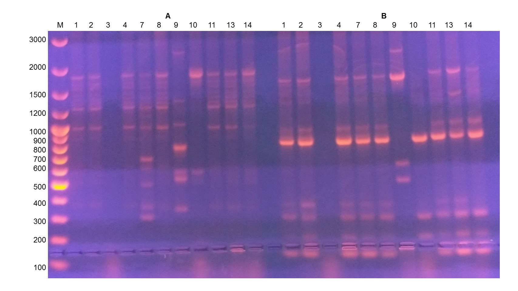
Genotyping B.subtilis strains using primer OPA-3 (left, A) and primer OPL-12 (right, B)
M - marker of DNA fragment length (GeneRuler 100 bp, Thermo Fisher Scientific)
Thus, these two primers can be successfully used for genotyping and identification (certification) of Bacillus subtilis strains, since they are capable to amplify several polymorphic DNA fragments for subsequent visualization.
4. Conclusion
1. The optimal temperature for genotyping B. subtilis using primers OPA-3 and OPL-12 is 37°C, the concentration of magnesium ions is 2.5 mM;
2. When genotyping B. subtilis strains, the short RAPD primers OPA-3 and OPL-12 were the most informative as compared with three other primers;
3. PCR genotyping of B. subtilis strains used as an active component in biopreparations allows certification of these strains to confirm their purity and genetic uniformity.