Changes in the Levels of Total Bile Acids in the Blood Serum of Minks with the Most Common Hepatopathies
Changes in the Levels of Total Bile Acids in the Blood Serum of Minks with the Most Common Hepatopathies
Abstract
Measurement of serum bile acid concentrations is a routine diagnostic test for assessing liver function. The main goal of this study is to evaluate the change in the level of serum bile acids in minks (Mustela vison Schreber, 1777) in the most common hepatotoxic conditions (toxic hepatitis, fatty hepatosis (fatty liver)). Analyzing the data of the study, we can highlight the following conclusions: in case of fatty hepatosis (fatty liver), the level of serum bile acids increased on average 2-2.5 times compared with the upper limit of the reference values, and there was no significant difference in the increase between males and females. In case of toxic hepatitis, a significant increase in the level of serum bile acids was noted, while in males this increase exceeds the upper limit of the reference intervals by 4–5 times, in females by 7–8 times. The indicators obtained in the study can later be used not only to determine the presence of hepatopathologies in animals, but also an attempt to differentiate them, which, together with classical biochemical indicators, will facilitate further diagnosis.
1. Introduction
According to most authors, throughout the history of human and veterinary medicine, bile acids (BA, cholic acids, cholenic acids) were thought to be, although undoubtedly unique molecules, but acting exclusively on the digestive tract and not noticeably influence on other organs. Ideas about their functions were narrowed mainly to lipids emulsification, control of intestinal secretion and local immunity. Lately with deeper knowledge about close interaction between different inner systems, elucidation of the BA functions has moved to a qualitatively new stage , , .
Analyzing BA levels in the blood serum is a routine diagnostic method for assessing liver function. BA are assembled in parenchymal liver cells from cholesterol. Cholic and chenodeoxycholic acids are the primary bile acids in most animals. After synthesis and prior excretion into bile, BA create complexes with amino acids – taurine and glycine. This reaction let the molecular weight of these fat-soluble compounds grow, making them more water-soluble, less reactive in an aqueous environment (bile in particular). Through an energy-dependent transport mechanism, these osmotically active steroid compounds pass the hepatocyte tubules walls and start the primary driving force for bile flow. BA are saved and concentrated in the gallbladder. In the bile, with an alkaline reaction, sodium and potassium salts of BA are formed, leading to better emulsifying properties. During meals, hormonal and neurohormonal factors envigorate the gallbladder contraction and the passage of BA into the duodenum. BA transform dietary fats into lipids and start metabolism of lipids and fat-soluble vitamins, and transport of water and electrolytes in the intestine. A dehydroxylation process occurs because of anaerobic microorganisms in the intestine, resulting in primary BA conversion into secondary ones. Thus, cholic acid is transformed into deoxycholic acid, and chenodeoxycholic acid – to lithocholic acid. Most BA get into portal circulation via active transport from the ileum, and less than 5% of the BA pool is excreted through the rectum. In general, BA are efficiently removed from the portal circulation during their first passage through the liver. As a result, only a slight rise in serum BA levels is recognised in healthy animals. After eating, enterohepatic BA circulation occurs several times , , , .
Currently, analyzing of BA levels in the blood is used as a marker of the functional state of the hepatobiliary system, however, it is said that such a method on itself doesn’t let differentiating the various etiology of shifts in liver function and should always be applied in combination with other methods for assessing liver function , , .
The main goal of this research is to rate levels of BA in blood serum in minks (Mustela vison Schreber, 1777) in the most widespread hepatotoxic conditions (toxic hepatitis, fatty hepatosis (fatty liver)).
2. Research methods and principles
Work were accomplished in 2022 at the Mermeriny fur farm (village Mermeriny, Kalinin district, Tver region). Palomino minks (Mustela vison Schreber, 1777) were chosen as model animals due to their specie's propensity for hepatopathy and the maximum rate of enterohepatic BA circulation among mammals. To study animals with both fatty hepatosis and toxic hepatitis, parity groups were created (10 females and 10 males in each, age – 1 year).
Before the experiment, the information from clinical examination carried out at the fur farm were analyzed, after which, based on the anamnesis and physical examination, candidates (10 females and 10 males, age – 1 year) with suspected fatty liver were selected. Subsequently, the presumptive diagnosis was confirmed on the basis of clinical and biochemical studies.
Due to the fact that toxic hepatitis is considered to be a widespread, but sporadically manifested pathology, clinically healthy animals (10 females and 10 males, 1-year-old) were selected to create a parity group for further induction of pathology in them. For randomization, a group (block) method with stratification by age and gender were applied. Animals were not stratified by weight and were randomly selected from the population . Toxic hepatitis was induced according to the method written in the Manual for the experimental (preclinical) study of new pharmacological substances, edited by Khabriev R.U. using ethylene chloride (1,2-dichloroethane), the toxicant dosage was calculated separately for each mink using interspecies dose conversion factors. According to the method, dichloroethane was diluted in olive oil to 50% solution, and was administered once orally at a dosage of 5 ml / kg . The manipulations performed as part of the experiment were allowed by the Committee of Bioethics of the FSBEI HE SPbSUVM, they correspond to the "European Convention for the Protection of Vertebrate Animals used for Experimental and Other Scientific Purposes", adopted in Strasbourg in 1987; and in accordance with Directive 2010/63/EU.
Total BA were identified using a BSBE bile acid kit (BSBE, China).
Results were statisticaly processed in the Statistica 6.0 software. Mean values of indicators (M), standard errors of means (±SEM) were calculated.
3. Main results
The BA level in the mink’s blood serum demonstrated in Figure 1. Absence of reliability calculation was due to the exploratory nature of the work and the absence of control groups.
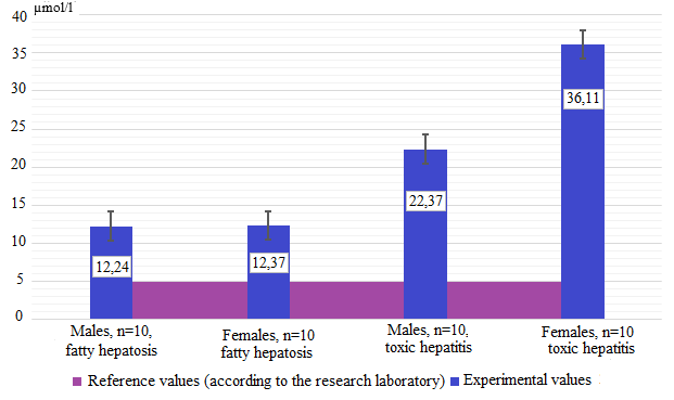
Figure 1 - Total bile acids level in the blood serum of the studied groups
With toxic hepatitis, a great rise in the level of serum bile acids was noted, in males this increase exceeds the upper limit of the reference intervals by 4–5 times, in females by 7–8 times. This contrast between males and females can be explained by the fact that the biochemical profile of females is more akin to cholesterol, a precursor of both bile acids and steroid hormones, which are always higher in the blood serum of females than in males.
Thus, shifts in BA levels described above relative to reference intervals that should be noted as metabolic parameters of hepatopathy in fur-bearing animals (in particular, minks). Further studies require confirmation with the help of morphological methods of research with the analysis of modern statistical methods.
4. Conclusion
Despite the fact that the predictive potential of bile acids in relation to the hepatobiliary system pathologies of various origins, although obvious, is not fully understood, some patterns between the corresponding pathology and changes in serum parameters can be determined.
The indicators obtained in the study can later be used not only to determine the presence of hepatopathologies in animals, but also an attempt to differentiate them, which, together with classical biochemical indicators, will facilitate further diagnosis.