Features of the Histological Structure of the Liver in Fatty Hepatosis in Minks
Features of the Histological Structure of the Liver in Fatty Hepatosis in Minks

Abstract
Fatty liver degeneration (fatty hepatosis) is extremely difficult to diagnose pathologies characterized by a course with no obvious clinical signs, especially in the early stages. Differential diagnosis in distinct from other hepatopathologies (in particular, from toxic and infectious hepatitis, amyloidosis and others) also causes significant difficulties, since comorbidity phenomena are observed at the initial stage of its formation, which is due to a single pathogenetic mechanism of their occurrence. The main goal of the study is to identify and analyze the main histological changes in the liver of minks with fatty liver (hepatosis).
Liver from sick animals was taken immediately after the planned slaughter according to the rules for the selection of pathological material.
Histological samples were made by standard methods with Van Gieson staining.
As a result of the study, the main histological signs of fatty degeneration of the liver were revealed: the pattern of the lobular structure is not expressed, the beam structure is smoothed out (the beam-radial structure of the lobules begins to fade against the background of moderately pronounced focal-diffuse large-drop fatty degeneration of hepatocytes); portal tracts with slight fibrosis, somewhat dilated, with lymphoid infiltration; large-drop infiltrative obesity of hepatocytes; bile ducts show cholestasis.
1. Introduction
Pathologies of the hepatobiliary system of various origins are one of the dominant pathologies in fur animals. Thus, according to some authors, in the Russian Federation up to 90% of animal mortality occurs from non-contagious diseases, among which the most significant is hepatosis (degenerative liver processes of non-infectious genesis) , , .
The main problem in the correct diagnosis for fatty hepatosis is its subclinical (latent) course at the initial stages of pathology. Differential diagnosis from other hepatopathologies also causes difficulties, since, the phenomenon of comorbidity is observed at the initial stage of its formation, which is due to a single pathogenetic mechanism of their occurrence , , .
To date, the diagnosis of fatty hepatosis presents significant difficulties, since it requires the exclusion of other liver diseases and the use of invasive examination methods. The study of clinical and biochemical markers of such liver damage has a low diagnostic value, since their changes are nonspecific and can occur in various pathologies of the hepatobiliary system , .
The most accurate way to diagnose and differential diagnosis of such pathologies is still considered to be a histological examination of damaged tissue, which is currently considered the so-called "gold standard" in determining the nature of hepatopathology. Morphological examination of liver biopsy specimens makes it possible to carry out a differential diagnosis of potential hepatopathy with high accuracy and, based on histological data, to predict the further course of the disease, as well as exclude other causes of liver damage , , .
The main goal of the study is to identify and analyze the main histological changes in the liver of minks with fatty liver (hepatosis).
2. Research methods and principles
The experiments were carried out in 2022 at the Mermeriny fur farm (village Mermeriny, Kalinin district, Tver region). Palomino minks (Mustela vison Schreber, 1777) were chosen as model animals.
Before the study, the results of clinical examination carried out at the fur farm were analyzed, after which, based on the anamnesis and physical examination, candidate animals (10 females and 10 males, age – 1 year) with suspected fatty liver were selected. Subsequently, the presumptive diagnosis was confirmed on the basis of clinical and biochemical studies. The clinical picture was similar to the symptoms of chronic fatty hepatosis: mild lethargy, reduced water intake, dull coat.
Liver from sick animals was taken immediately after the planned slaughter according to the rules for the selection of pathological material .
Histological samples were made by standard methods with Van Gieson staining . The van Gieson stain uses a mixture of picric acid and acid fuchsin. This stain is the simplest method for differentiating collagen fibers from other connective and smooth muscle components. Collagen fibers are stained red, and cells of smooth muscle and striated tissue, keratinizing stratified squamous epithelium and hyaline in different shades of yellow up to brownish, the cell nucleus in black. For a complete study of the histological structure, the sections were examined at the following magnifications: x10x10, x10x20, x10x40, x10x100.
Morphometric studies and their statistical processing were carried out using the ImageJ open source software.
3. Main results
The results of histological examination of the mink liver with fatty hepatosis are shown in Figures 1-4. The histological picture was similar in all studied samples, which was confirmed by the "ImageJ" software.
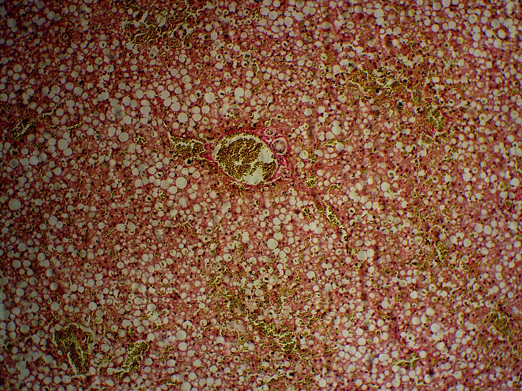
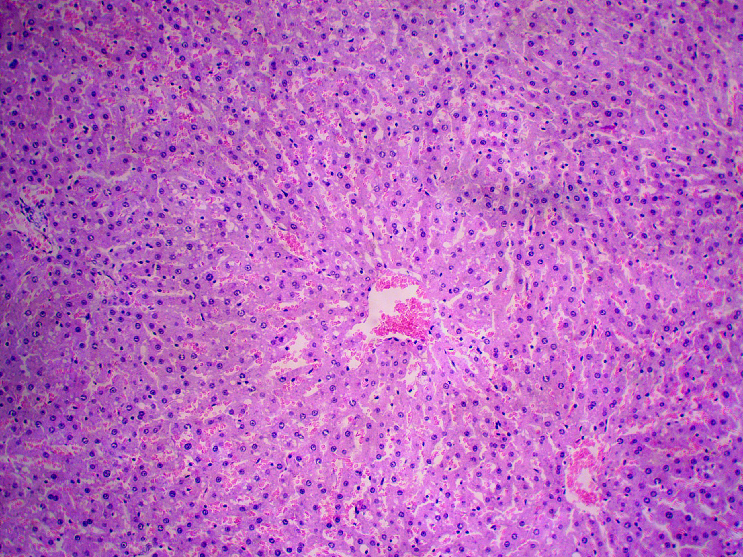
Histological picture of the mink liver with fatty hepatosis
Van Gieson stain, magnification x10x10
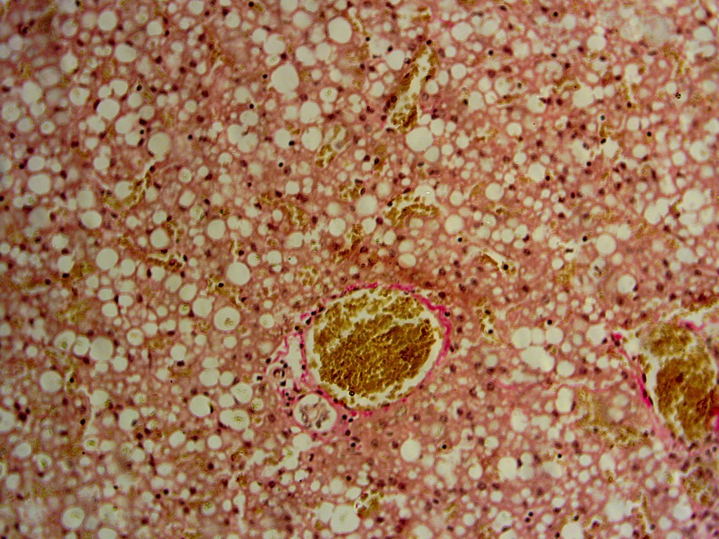
Histological picture of the mink liver with fatty hepatosis
Van Gieson stain, magnification x10x20
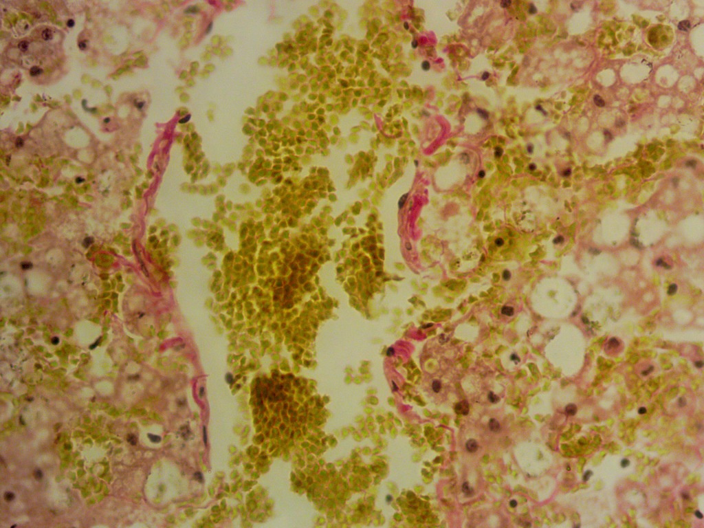
Histological picture of the mink liver with fatty hepatosis
Van Gieson stain, magnification x10x40
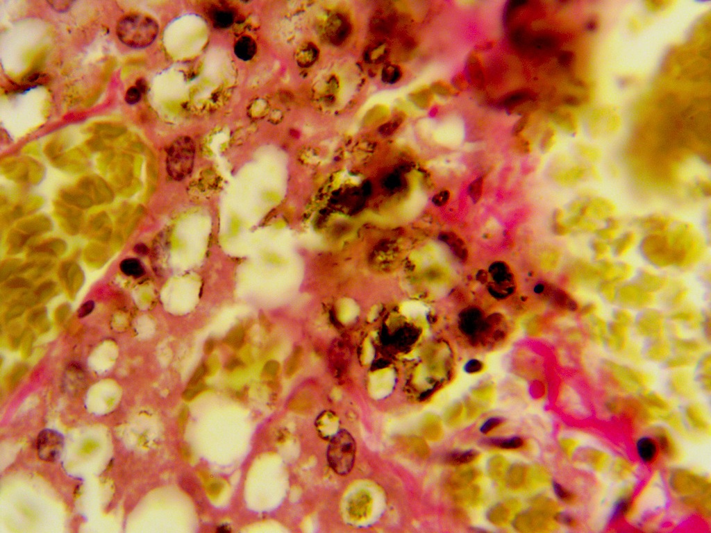
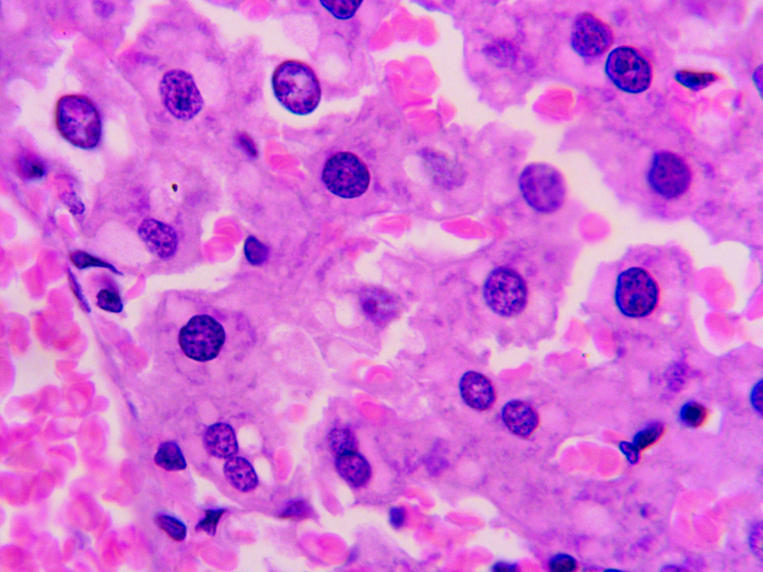
Histological picture of the mink liver with fatty hepatosis
Van Gieson stain, magnification x10x100
Three structural-functional units can be distinguished in the organization of the liver:
1) hepatic lobule – a hexagonal prism through the center of which the central vein passes, collecting blood from the sinusoidal capillaries. Next to the lobule is a portal tetrad, which consists of an interlobular artery (a hepatic artery branch of the systemic circulation), an interlobular vein (the portal vein branch), an interlobular bile duct (into which bile flows from the bile capillaries of the lobule) and an interlobular lymphatic vessel;
2) the portal lobule, which includes segments of three neighboring classical hepatic lobules surrounding the triad. Therefore, it has a triangular shape, a triad lies in its center, and on the periphery, in the corners, veins (central). In this regard, in the portal lobule, the blood flow through the blood capillaries is directed from the center to the periphery;
3) hepatic acinus – part of the tissue that surrounds the portal triad and includes lymphatic vessels, nerve fibers and adjacent sectors of two or more lobules. One acinus contains about 20 hepatic cells located between the portal triad and the central vein of each lobule. The acinus microvascular frame consists of the above blood and lymphatic vessels, sinusoids and nerves. It is the microcirculatory unit of the liver.
However, histological examination of the mink liver revealed polygonal-shaped hepatocytes, predominantly mononuclear (multinuclear and polyploid hepatocytes reflect adaptive changes in the liver, since these cells are able to perform their functions much more intensively than ordinary hepatocytes). The pattern of the lobular structure is not pronounced, the beam structure is smoothed out (the beam-radial structure of the lobules begins to fade against the background of a moderately pronounced focal-diffuse large-drop fatty degeneration of hepatocytes). Sinusoids, Kupffer cells are well visible. Severe venous-capillary plethora of the liver vessels. Portal tracts with slight fibrosis, somewhat dilated, with lymphoid infiltration. Large-drop infiltrative obesity of hepatocytes. Interlobular bile ducts of the liver have a different diameter. Depending on the diameter, they are lined with a single-layered cubic or prismatic epithelium. There is cholestasis in the bile ducts. In hepatocytes and Kupffer cells, there are deposits of hemosiderin (dark yellow pigment, with the chemical structure based on iron oxide, formed during the breakdown of hemoglobin and subsequent denaturation and deproteinization of the ferritin protein responsible for storing iron in the body). Hemosiderin in the liver indirectly points to chronic intoxication.
The histological picture of the structure of the mink liver is characteristic of severe fatty degeneration of the liver (based on the fact that the mink is grown artificially, of alimentary origin). For comparison, histological sections of the mink liver from the same fur farm, but without clinical signs of hepatopathology, are given. The liver was taken during the planned slaughter of animals (Figure 5, 6).
Histological picture of the liver of healthy minks
Hematoxylin-eosin stain, magnification x10x10
Histological picture of the liver of healthy minks
Hematoxylin-eosin stain, magnification x10x100
4. Conclusion
One of the main problems in addressing the issues of prevention and treatment of hepatopathy in fur-bearing animals is the lack of a relevant diagnostic base for the timely detection of this type of pathology. Most liver diseases go latently or with non-specific symptoms, with the rapid development of pathological processes. The situation with diagnostic approaches is most indicative of the fact that the method for diagnosing pathologies of the hepatobiliary system, based on needle biopsy, which has become established as the "gold standard", is still the most representative and informative.
Pathological anatomical signs of hepatosis, confirmed by histological examination, allow timely detection of the disease and taking the necessary measures to treat animals and eliminate the causes of hepatosis.